Sabtu, 16 Juni 2012

VIRTUAL MACHINE

VIRTUAL MACHINE

 A. Sejarah dan Definisi

 Mesin virtual pada mulanya didefinisikan oleh Gerard J. Popek dan Robert P. Goldberg pada tahun 1974 sebagai sebuah duplikat yang efisien dan terisolasi dari suatu mesin asli. Pada masa sekarang ini, mesin-mesin virtual dapat mensimulasikan perangkat keras walaupun tidak ada perangkat keras aslinya sama sekali.
Contoh, program yang ditulis dalam bahasa Java akan dilayani oleh Java Virtual Machine (JVM) dengan cara memberikan perintah-perintah yang dimengerti JVM yang selanjutnya akan memberikan hasil yang diharapkan. Dengan memberikan layanan seperti ini kepada program tersebut, perangkat lunak JVM ini berlaku sebagai sebuah "mesin virtual", sehingga program tidak lagi perlu untuk mengakses langsung melalui sistem operasi ataupun perangkat keras yang sangat bervariasi dan memerlukan pemrograman masing-masing secara spesifik.
Mesin virtual terdiri dari dua kategori besar, dipisahkan menurut cara penggunaan dan tingkat keterhubungannya dengan mesin-mesin aslinya. Sebuah mesin virtual sistem adalah perangkat yang berupa platform sistem yang lengkap dan dapat menjalankan sebuah sistem operasi yang lengkap pula. Sebaliknya, mesin virtual proses didesain untuk menjalankan sebuah program komputer tertentu (tunggal), yang berarti mesin virtual ini mendukung proses tertentu juga. Karakteristik mendasar dari sebuah mesin virtual adalah batasan-batasan bagi perangkat lunak yang berjalan di dalam mesin tersebut, sumber daya yang dibatasi, dan tidak dapat mengakses ke luar tembok batasan dunia maya itu.

B. Kegunaan

Teknologi virtual  machine memiliki  banyak kegunaan seperti  memungkinkan konsolidasi  perangkat keras,  memudahkan  recovery  sistem,  dan menjalankan perangkat lunak terdahulu.  Salah  satu penerapan penting dari teknologi  VM adalah   integrasi   lintas  platform. Beberapa   penerapan   lainnya   yang   penting adalah:
1.    Konsolidasi  server
Jika beberapa server menjalankan aplikasi yang hanya memakan sedikit sumber daya, VM dapat digunakan untuk menggabungkan aplikasi-aplikasi tersebut sehingga berjalan pada satu server saja, walaupun aplikasi tersebut memerlukan sistem operasi yang berbeda-beda.
2.    Otomasi dan konsolidasi lingkungan pengembangan dan testing
Setiap VM  dapat   berperan   sebagai   lingkungan   yang   berbeda,   ini  memudahkan pengembang sehingga tidak perlu menyediakan lingkungan tersebut secara fisik.
3.    Menjalankan perangkat  lunak terdahulu
Sistem operasi dan perangkat lunak terdahulu dapat dijalankan pada sistem yang lebih baru.
4.    Memudahkan  recovery  sistem
Solusi   virtualisasi   dapat   dipakai   untuk rencana recovery sistem yang memerlukan portabilitas dan fleksibilitas antar platform.
5.    Demonstrasi perangkat lunak
Dengan teknologi VM, sistem operasi yang bersih dan konfigurasinya dapat disediakan secara cepat.

C. Kelebihan dan kekurangan

  Kelebihan :
 1. Hal  keamanan.
     VM memiliki  perlindungan yang  lengkap pada berbagai sistem  sumber   daya,   yaitu   dengan  meniadakan   pembagian   sumber   daya secara  langsung,  sehingga  tidak ada masalah proteksi  dalam VM.  Sistem VM adalah kendaraan yang sempurna untuk penelitian dan pengembangan sistem operasi. Dengan VM, jika terdapat suatu perubahan pada satu bagian dari mesin, maka dijamin tidak akan mengubah komponen lainnya.
  2. Memungkinkan   untuk  mendefinisikan   suatu   jaringan   dari   Virtual Machine   (VM).
Tiap-tiap   bagian  mengirim  informasi  melalui   jaringan komunikasi  virtual.  Sekali   lagi,   jaringan  dimodelkan   setelah komunikasi fisik jaringan diimplementasikan pada perangkat lunak.

Kekurangan:
 Beberapa kesulitan utama dari konsep VM, diantaranya adalah:
1. Sistem penyimpanan.
Sebagai contoh kesulitan dalam sistem penyimpanan adalah   sebagai   berikut:   Andaikan   kita   mempunyai   suatu   mesin   yang memiliki  3  disk drive  namun  ingin mendukung 7 VM.  Keadaan  ini   jelas tidak memungkinkan bagi kita untuk dapat mengalokasikan setiap disk drive untuk  tiap VM,  karena perangkat   lunak untuk mesin virtual   sendiri  akan membutuhkan   ruang disk   secara   substansial  untuk menyediakan  memori virtual  dan  spooling.  Solusinya   adalah dengan menyediakan disk  virtual atau   yang   dikenal   pula   dengan  minidisk,   dimana   ukuran   daya penyimpanannya   identik   dengan   ukuran   sebenarnya.   Dengan   demikian, pendekatan VM juga menyediakan sebuah antarmuka yang identik dengan perangkat keras yang mendasari.
2. Pengimplementasian sulit.
Meski konsep VM cukup baik, namun VM sulit diimplementasikan.
Contoh virtual machine : Vmware, Xen VMM , Java VM

Jenis-jenis dari VM adalah:
1.    VM sistem di mana sebuah VM dapat menjalankan sebuah  sistem operasinya  sendiri.
2.    VM proses  di  mana VM hanya menjalankan sebuah proses saja.

Kemudian VM juga dibagi berdasarkan tingkat virtualisasinya:
1.    Virtualisasi   penuh   yang  mensimulasikan   seluruh   fitur  perangkat  keras   sehingga memungkinkan perangkat   lunak berjalan pada VM tanpa  modifikasi.
2.    Virtualisasi   paruh,   di  mana   tidak   semua   fitur perangkat keras disimulasikan.
3.    Virtualisasi asli, yang mana merupakan   virtualisasi   penuh   yang   digabungkan   dengan   bantuan   perangkat keras yang mendukung virtualisasi.


efek 2 warna


Download Photoshop asli klik Disini

poster kesehatan


Download corel draw asli klik Disini

Kamis, 17 Mei 2012

Determination of Hematocrit Value


Hematocrit or erythrocyte volume of the compressed (packed cell volume, PCV) is the percentage volume of erythrocytes in the blood is compressed by playing at a certain speed and in a certain time. The purpose of this test is to determine the concentration of erythrocytes in the blood.

Based on the reproducible and simple, this examination is the most trustworthy among other checks, namely hemoglobin and erythrocyte count. Can be used as a simple filter test against anemia.

Hematocrit or PCV values ​​can be set using the automatic hematology analyzer or manually. Manually hematocrit measurement method known there are two, namely :

Methods makrohematokrit
At the macro method, as much as 1 ml blood samples (EDTA or heparin blood) were included in Wintrobe tubes measuring 110 mm long with a diameter of 2.5-3.0 mm and 0-10 mm scale. The tubes then were centrifuged for 30 minutes at 3,000 rpm. High erythrocyte column is the hematocrit value is expressed in%.

Methods mikrohematokrit
At the micro method, blood samples (capillary blood, blood EDTA, heparin blood or blood-potassium-ammonium oxalate) is inserted in a capillary tube having a length of 75 mm in diameter and 1 mm. Capillary tubes are used there are two kinds, namely containing heparin (marked red) for capillary blood samples (direct), and without anticoagulant (marked blue) blood to EDTA / heparin / ammonium-potassium-oxalate.

Examination procedures are: blood sample is inserted into the capillary tube until 2 / 3 the volume of the tube. One end of the tube is closed with putty (clay) and then centrifuged for 5 minutes at 15,000 rpm. High erythrocytecolumn were measured with a hematocrit reader, its value expressed in%.

Mikrohematokrit method is more widely used because in addition to quite a short time, blood samples required is also small and can be used for samples without anticoagulant that can be obtained directly.

Reference Values
Adult male : 40 - 52%
Adult women : 35-47%
Newborns : 44-72%
Children aged 1-3 years : 35-43%
Children aged 4-5 years : 31-43%
Children aged 6-10 years : 33-45%

Clinical Problems
Decreased levels: acute blood loss, anemia (aplastic, hemolytic, folic acid deficiency, pernicious, sideroblastik, sickle cell), leukemia (lymphocytic, myelogenous, monositik), Hodgkin's disease, limfosarkoma, organ malignancies, multiple myeloma, cirrhosis of the liver, protein malnutrition , deficiency of vitamins (thiamine, vitamin C), gastric or duodenal fistula, peptic ulcer, failure, chronic kidney, pregnancy, SLE. The influence of drugs: antineoplastic, antibiotics (chloramphenicol, penicillin), radioactive medicine.

Increased levels: dehydration / hypovolemia, severe diarrhea, polycythemia vera, eritrositosis, diabetic acidosis, pulmonary emphysema late stage, whereas cerebral ischemia, eclampsia, surgery, burns.

Factors that may affect the laboratory's findings :
If a blood sample taken at the arm that is attached intra-venous lines, tend to be low hematocrit values ​​due to hemodilution. Installation of rope tourniquet that is too long the potential to cause hemoconcentration, so that the hematocrit value can be increased. Capillary blood sampling: punctures lacking in so that the volume gained slightly and squeeze the blood must be squeezed-out, which pierced the skin is still damp with alcohol so that the diluted blood, clots occur in the blood drops because of slow work.

Hematology test, Osmotic fragility erythrocytes

When erythrocytes are in solution hipotonis, osmolalitasnya fluid levels lower than normal plasma or serum (less than 280 mOsm / kg)

Erythrocyte osmotic fragility test (also called erythrocyte osmotic resistance) is done to measure the ability to withstand the occurrence of erythrocyte hemolysis (destruction of red blood cells) in asolution that hipotonis. The trick is as follows: erythrocytes were dissolved in saline solution with various concentrations. If hemolysis occurs at a slightly saline solution hipotonis, this state is called an increase in erythrocyte fragility (= reduction in resistance / durability erythrocytes), and if hemolysis occurs at a very hipotonis salinesolution, this situation indicates decreased osmotic fragility (= increase in erythrocyte resistance).

Hemoglobin out of the cells in each tube containing a solution of NaCl of different levels. Hb then determined fotokolorimetrik. The results are reported in percentages (%) hemolysis. Collection of hemolysis results are plotted in a curve as compared with normal erythrocytes data. In the state of increased fragility, erythrocytes are usually spherical, and the curves seem to shift to the right. While the decrease in fragility, thin and flat-shaped erythrocytes, the curve appears shifted to the left.

Clinical Problems
DECREASE fragility: Thalassemia major and minor (Mediterranean anemia or Cooley's anemia), anemia (iron deficiency, folic acid deficiency, vitamin B6 deficiency, sickle cell) hemoglobin C disease, polycythemia vera, post splenectomy, acute liver necrosis and sub-acute, jaundice obstructive.

IMPROVEMENT fragility: hereditary spherocytosis, transfusions (ABO and Rhesus incompatibility), autoimmune hemolytic anemia (AIHA), hemoglobin C disease, drug toxicity or chemicals, chronic lymphocytic leukemia, burns (thermal).

Procedure
This test is usually performed on fresh blood samples of less than 3 hours and / atu 24-hour blood samples were incubated at 37 ° C. Blood samples used in the form of heparin blood or blood "defibrinated". There are no restrictions on food or beverage intake.

In this test made solution with different concentrations of NaCl. Assessment of results with fotokolorimetri method (using photometer or spectrophotometer).

Prior to testing, provide a first buffer stock solution of NaCl 10% made from 9 grams NaCl, 1.365 g Na2HPO4, and 0.215 grams NaH2PO4.H2O. The material is then diluted with distilled water to 100 ml. Before being used for inspection, make asolution of 1.0% NaCl principal by dissolving 5.0 ml of saline buffer stock of 10% with distilled water to 50.0 ml. Next do the testing as follows :
  1. Provide 12 pieces and then make a dilution tube multilevel solution of NaCl concentration: 0.85%, 0.75%, 0.65%, 0.60%, 0.55%, 0.50%, 0.45%, 0, 40%, 0.35%, 0.30%, 0.20% and 0.10%, respectively of 5.0 ml. NaCl solutions were made from the basicsolution of NaCl 1.0%.
  2. Add into the canisters each 50 mL blood sample. Mix (homogenization) by way of tossing and turning the tube several times.
  3. Inkubasikan for 30 minutes at room temperature.
  4. Mix (homogenization) again and then worry about (centrifuges) of each tube for 5 minutes at 3000 rpm.
  5. Measure the absorbance (OD) of the supernatant at λ 540 nm with the blank tube to supernatant-1 (0.85% NaCl).
  6. Calculate the% hemolysis by dividing the absorbance (OD) of samples with the absorbance (OD) tubing to 12 multiplied by 100%.
  7. Create a curve with the concentration of NaCl as the axis (x) and% hemolysis as the ordinate (y). Compare with the curve of normal blood controls.

Normal Value
The beginning of hemolysis on the concentration of NaCl 0.40% - 0.45%
Complete hemolysis at a concentration of 0.30% NaCl - 0.35%

The percentage of hemolysis in normal circumstances are :
97-100% hemolysis in 0.30% NaCl
50-90% hemolysis in 0.40% NaCl
5-45% hemolysis in 0.45% NaCl
0% hemolysis in 0.55% NaCl

Factors Affecting Laboratory Findings
  • plasma pH, temperature, glucose concentration, and oxygen saturation in the blood
  • Long-lived erythrocytes tend to have high osmotic fragility
  • Blood samples taken over 3 hours can show increased osmotic fragility.
Source of the article and continue reading : http://labkesehatan.blogspot.com

Taking Blood Veins With Vacuum Tubes, Vacutainer


bd.com
Vacuum tube was first marketed by U.S. company BD (Becton-Dickinson) under the trade name Vacutainer. This type of tube in the form of a vacuum tube, made of glass or plastic. When the tube is attached to the needle, the blood will flow into the tube and the flow stops when a certain volume has been reached.

Needles used consists of two needles connected by a threaded connection. The needle on the anterior side is used to puncture the vein and the needle on the posterior side of the tubes plugged. Posterior needle encased by the material of rubber so as to prevent the blood from the patient flows out. Threaded connection serves to attachthe needle in a holder and ease when pushing the needle tube mounted directly on the posterior.

The advantage of using this retrieval method is, no need to divide the sample into several tubes of blood. Just one stabbing, can be used for multiple tubes alternately according to the type of tests required. For the purposes of the bacteria culture test, thismethod is also better because the patient's blood can flow directly into the tube containing the bacteria culture media. Thus, the possibility of contamination duringsample transfer to the decision by way of the manual can be avoided.

The drawback difficult decision in the elderly, small children, infants, or if the vein is unreliable (small, fragile), or if the patient is obese. To overcome this may be used needle with wings (winged needle).

Winged needle or needles is often also called "butterfly" needle vakutainer nearly equal as mentioned above. The difference is, betweenthe needle anterior and posterior wings there are two pieces of plastic at the base of the anterior needle and the needle tube that connects the anterior and posterior. If the proper insertion of the vein, blood will look into the hose (flash).

Procedure :
  1. Prepare the necessary tools: needles, cotton 70% alcohol, a hedge strap (tourniquet), plaster, vacuum tubes.
  2. Attach the needle to the holder, make sure it is firmly.
  3. Approach the patient in a calm and friendly; try to patients as comfortable as possible.
  4. Identification of patients correctly according to the data at the request sheet.
  5. Verification of the patient, such as fasting or consumption of drugs. Record if the patient is taking certain medications, etc. are not fasting.
  6. Ask the patient to straighten his arm, select the arm that did a lot of activity.
  7. Have the patient make a fist.
  8. Attach a rope hedge (tourniquet) is approximately 10 cm above the elbow fold.
  9. Select the median cubital or cephalic vein. Do palpability (palpation) to ensure the position of the vein; vein palpable as a small pipe, elastic and has a thick wall. If the veins are not palpable, do the sorting of the wrist to the elbow, or warm compresses for 5 minutes the arm.
  10. Clean the skin on the parts to be taken with alcohol 70% cotton and let dry. Skin that has been cleaned do not hold anymore.
  11. Puncture the vein with a needle hole position facing up. Insert the tube into the holder and push that needle stuck in the posterior part of the tube, then the blood will flow into the tube. Wait until the blood stops flowing. If you require multiple tubes, having first filled the tube, remove and replace with a second tube, and so on.
  12. Remove the tourniquet and ask the patient to open his fist. The volume of blood taken approximately three times the amount of serum or plasma required for the examination.
  13. Put the cotton on the injection site and then immediately release / pull the needle. Press the cotton and plasters couple sat for about 15 minutes. Do not pull the needle before the tourniquet was opened.
Accommodate Blood In Tube
Some types of blood sample tubes used in the clinical laboratory practices are as follows :
Tube red cap. This tube without the addition of additives, the blood will be frozen and the serum separated by centrifugation. Commonly used for examination of blood chemistry, immunology, serology and blood bank (crossmatching test)

Tube yellow cap. It contains a gel separator tubes (serum separator tube / SST) whose function is to separate serum and blood cells. After centrifugation, the serum will be at the top of the gel and the blood cells are under the gel. Commonly used forexamination of blood chemistry, immunology and serology

Tube light green lid. It contains a gel separator tube (plasma separator tube / PST) with lithium heparin anticoagulant. After centrifugation, the plasma will be at the top of the gel and the blood cells are under the gel. Commonly used forexamination of blood chemistry.

Purple or lavender tube cap. This tube contains EDTA. Commonly used for complete blood count and blood banks (crossmatch)

Tube blue cap. This tube contains sodium citrate. Commonly used for examination of coagulation (eg PPT, APTT)

Tube green cap. This tube contains sodium or lithium heparin, commonly used for the examination of erythrocyte osmotic fragility, blood chemistry.

Dark blue cap tube. This tube contains EDTA-free metals, generally used for examination of trace elements (zinc, copper, mercury) and toxicology.

Tube light gray cap. This tube contains sodium fluoride and potassium oxalate, were used for examination of glucose.

Black cap tube; containing sodium citrate buffer, used for checking the LED (ESR).

Pink cap tube; containing potassium EDTA, used for examination imunohematologi.

White cap tube; potassium EDTA, used for examination of molecular / PCR and bDNA.

Tube yellow with a black cap at the top; containing culture media, used for microbiological examination - aerobic, anaerobic and fungal

Accommodate some of the important things in the blood sample is :
Blood from syring or injections should be inserted into the tube by removing the needle and the blood flow slowly through the tube wall. Enter the blood by way of spraying, especially without removing the needle, potentially causing hemolysis. Blood entering into the vacuum tube by a needle on the cap tube, allow blood to flow until it stops itself when the volume has been met.

Homogenization of samples if using anticoagulants by twisting the tube 4-5 times or tossing and turning the tube 5-10 times by softly. Whisk thesample potentially cause hemolysis.

The order to enter the blood samples into vacuum tubes are: first - bottle culture (culture) of blood or yellow-black cap tube second - coagulation tests (tube blue cap), third - non-additive tube (red cap), the fourth - the red cap tubes or yellow with separator gel or clot activator, tube cap purple / lavendet (EDTA), green cap tube (heparin), gray cap tubes (NaF and Na oxalate).

Image source : bd.com, Thanks, references from my teachers address : labkesehatan.blogspot.com

Protein C-reactif

Protein C-reactif (C-reactive protein, CRP) are made by the liver and secreted into the bloodstream. CRP circulates in the blood for 6-10 hours after the acute inflammatory process and tissue destruction. Levels peaked within 48-72 hours. As with any test erythrocyte sedimentation rate (erithrocyte sedimentation rate, ESR), CRP is a non-specific test but the existence precedes the CRP increase during inflammation and necrosis of the LED and then immediately return to normal levels.

CRP is one of several proteins that are often referred to as acute phase proteins and used to monitor changes in the acute inflammatory phase is associated with many infectious diseases and autoimmune diseases. Some circumstances in which CRP may increase was found arthritis (rheumatoid arthritis), rheumatic fever, breast cancer, colitis, inflammatory disease stage (pelvic inflammatory disease, PID), Hodgkin's disease, SLE, bacterial infections.

CRP is also elevated in the final trimester of pregnancy, the use of intrauterine contraceptive devices and oral contraceptive drug effect.

CRP test is often performed repeatedly to evaluate and determine whether the treatment is effective. CRP is also used to monitor wound healing and to monitor post-surgical patients, organ transplant, or burns as an early detection system for possible infections.

High-sensitive CRP (hs-CRP)
This test can detect the inflammation that occurs due to the formation of atherosclerotic plaque in coronary arteries. hsCRP is a highly sensitive laboratory test for the risk of cardiovascular disease. This test is often done in conjunction with the test lipid profile (cholesterol, triglycerides, HDL, LDL). Positive hsCRP value is much lower than the standard value of serum CRP so that it becomes more useful test in detecting the risk of coronary heart disease (coronary heart disease, CHD), stroke, and peripheral arterial disease.

American Heart Association and the U.S. Centers for Disease Control and Prevention have defined risk groups as follows :

  • Low risk: less than 1.0 mg / L
  • Average Risk: 1.0 to 3.0 mg / L
  • High risk: above 3.0 mg / L

Those values ​​are only a part of the evaluation process for kardiovaskuler.Tambahan disease risk factors to consider are the elevated levels of cholesterol, LDL, triglycerides, and glucose. In addition, smoking, high blood pressure (hypertension), and diabetes also raise the level of risk.

Procedure
CRP test can be performed manually using agglutination methods or other more advanced methods, such as sandwiches imunometri. Agglutination tests carried out by adding the latex particles coated with anti-CRP antibodies in the serum or plasma agglutination patient so that it will happen. To determine the titer of CRP, serum or plasma diluted with buffer glycine patients with multilevel dilution (1 / 2, 1 / 4, 1 / 8, 1 / 16 and so on) and then reacted with latex. CRP titer is the highest dilution that still occur agglutination.

Imunometri sandwich tests performed by measuring the intensity of colors using Nycocard Reader. Consecutive samples (serum, plasma, whole blood) and conjugate dropped on the coated membrane antibody testsspecific mononklonal CRP. CRP in the sample captured by antibodies bound to colloidal gold particle conjugates. Free conjugate was washed with wash solution (washing solution). If there is at the level of CRP in pathological samples, it will form a red-brown color in the test area with color intensity proportional to the content. Color intensity was measured quantitatively using a reader NycoCard II.

Normal reference value of CRP with imunometri sandwich method is <5 mg / L. This reference value would be different depending on each laboratory reagents and methods used.

hepatitis A (HAV)

What is hepatitis A and how is it transmitted ?
Hepatitis A is caused by the hepatitis A virus (HAV). HAV is spread through food / drink contaminated with fecal matter (stool) of an infected person enter another person's mouth. HAV is mainly transmitted through raw or inadequately cooked, handled or prepared by someone withhepatitis A (although maybe he did not know he was infected).

Drinking water or ice that is contaminated with feces is another source of infection, as well as shellfish that are not cooked enough. HAV can be transmitted through "rimming" (oral-anal sex, or between the mouth and anus). HAV is rarely transmitted through blood-to-blood.

Hepatitis A is an acute form of hepatitis, meaning do not cause chronic infection. Once we've had hepatitis A, we can not be infected again. However, we can still be infected with other hepatitis viruses.

What are the symptoms of hepatitis A?
Not all people infected with HAV will have symptoms. For example, many infants and young children infected with HAV do not experience any symptoms. Symptoms are more likely to occur in older children, adolescents and adults.

Symptoms of hepatitis A (acute hepatitis and in general) can including :
  • skin and whites of the eyes become yellow (jaundice)
  • Fatigue
  • right-upper abdominal pain
  • Loss of appetite
  • Weight loss
  • Fever
  • Nausea
  • Diarrhoea or diarrhe
  • Vomiting
  • Water arts such as tea and / or dirt-colored putty
  • joint pain

HAV infection can also increase the levels of enzymes made by the liver to be above normal in the blood. The immune system require up to eight weeks to remove the HAV from the body. If symptoms develop, generally within two to four weeks after infection. Symptoms ofhepatitis A is generally only one week, but can be more than a month. Approximately 15 percent of people with hepatitis A have symptoms from six to nine months. Approximately one in 100 people infected with HAV may experience rapid and severe infections (so-called 'fulminant'), which - very rarely - can lead to liver failure and death.

How is hepatitis A diagnosed ?
Diagnosis of hepatitis A is confirmed by blood tests. The doctor will order these tests if we are experiencing symptoms of hepatitis A or if we want to know whether you were infected with HAV in the past. The blood test looks for two types of antibodies to the virus, which referred to as IgM and IgG (Ig stands for immunoglobulin). First, look for IgM antibodies, produced by the immune system of five to ten days before symptoms appear and usually disappear within six months. Tests also search for IgG antibodies, IgM antibodies are replaced and protect against HAV infection.

  • When blood tests show negative for IgM antibodies and IgG, we probably have never been infected with HAV, and should consider getting vaccinated against HAV.
  • If you are positive for IgM antibodies and negative to IgG, our chances of contracting HAV in six past month, and the immune system is being removing a virus or infection is becoming increasingly severe.
  • When the tests showed negative for IgM antibodies and positive for IgG antibodies, we may be infected with HAV in an earlier time, or we already vaccinated against HAV. We are now immune to HAV.

Note : Hepatitis A is endemic in Indonesia. This means that the majority of Indonesian people ever exposed to HAV during childhood, and most likely will be immune to infection again. Because of this, most doctors HAV test is not considered beneficial for people with HIV in
Indonesia.

How to people with HIV ?
People with HIV do not have the risk of becoming infected with HAV higher
than others. However, several studies indicate that people with HIV is more likely to experience symptoms of hepatitis A for Longer term, the meaning may take longer to recover fully from hepatitis A. One other important issue to consider is that many HIV-positive people taking antiretroviral drugs can be bad for the liver. Some of these drugs can worsen the symptoms ofhepatitis A. Because of this, maybe we should stop using all drugs so that the hepatitis A began to recover or liver enzyme levels returned to normal. Talk with your doctor before any medication dismiss.

How is hepatitis A treated ?
The usual treatment for hepatitis A is a break at sleep. There are also important to drink plenty of fluids, especially when we experiencing diarrhea or vomiting. Painkillers which counter, such as ibuprofen can reduce the symptoms hepatitis A, but we should talk more check with your doctor.

When we feel we may have been exposed to HAV - for example when someone in our household recently been diagnosed with hepatitis A - we should see a doctor to discuss the benefits of immune globulin injection (also called as gamma globulin). Immune globulin contains lots of antibodies against HAV, which can help prevent the onset of disease when we are exposed to the virus. Immune globulin must be given within two to six weeks after we might have been exposed to HAV. When we receive immune globulin to preventhepatitis A, we should also receive hepatitis A vaccine (discussed below).

How can hepatitis A be prevented ?
The best way to prevent hepatitis A is vaccination. Vaccination requires two injections, usually given with a time gap of six months. Side effects of hepatitis A vaccination, if it occurs, is usually mild and may include soreness at the injection site and mild flu-like symptoms. Also available is a combination vaccine for viralhepatitis A and B. HAV vaccine is very effective - more than 99 percent of people who receive a vaccination immune to viruses and not be exposed tohepatitis A if exposed. There is little doubt that HAV vaccination in people with very low CD4 counts may not provide immunity (due to very weak immune systems), so it should be vaccinated at a CD4 count is still quite high.

When we feel we have not been infected with hepatitis A, we should discuss with your doctor. Because people with HIV often experience more severe symptoms when infected with HAV, and our heart is essential to remove the remaining end of ARV drugs, HAV vaccination is recommended for people with HIV. Vaccination is especially important for people with HIV and hepatitis B or C.

Although we have not received vaccination against hepatitis A, there are some things we can do to prevent
HAV infection:
  • Avoid water, including ice, which could be contaminated with feces
  • Avoid raw shellfish or undercooked
  • Always wash hands with soap and water after using the room
  • bathing, changing diapers and before preparing
  • or eating food
  • Wearing a latex barrier ('dental dam') to sex oralanal

Anti-cardiolipin Antibodies (ACA) Test

Anticardiolipin Antibodies are proteins found in the body that works against kardiolipin. Kardiolipin and other related phospholipids are lipid molecules that are usually found in cell membranes and platelets as well as having an important role in regulating blood clotting. Whenantibodies produced against kardiolipin, they will increase the risk of blood clots forming an undue (thrombosis) in the arteries and veins.

Anticardiolipin Antibodies belong to a group of antiphospholipid antibodies (APA) in conjunction with lupus anticoagulan (LA). LA causes the elongation of activated partial thromboplastin time (APTT). Clinical manifestations associated with the respective antibody appeared similar, namely thrombosis. Clinical experience suggests that venous thrombosis may be associated with LA, and arterial thrombosis is more likely associated with a high titer of ACAantibodies. Antiphospholipid antibodies are acquired abnormalities; may occur in association with systemic autoimmune disorders or by itself. Patients remain at risk for thrombosis as long as there are autoantibodies.

Clinical Problems
Elevated levels of anticardiolipin antibodies found in antiphospholipid syndrome (thrombosis of arteries and veins are recurrent, recurrent miscarriage), autoimmune diseases (SLE, HIV / AIDS), preterm labor, pre-eclampsia, intrauterine growth retardation, thrombocytopenia, malignancy (leukemia, lymphoproliferative disorders and plasmasitik, solid tumors), infections (bacteria, viruses, protozoa), neurologic events including transient ischemic attack and stroke, liver disease, and disease dermatologik (livedo reticularis, acrocyanosis, pyoderma, extensive skin necrosis). The influence of drugs: chlorpromazine, procainamide, kuinidin, penicillin, many antibiotics, phenytoin.

Procedure
Anticardiolipin antibodies consists of three kinds, namely IgM, IgG, and IgA. Tests for anticardiolipin IgM and IgG antibodies are often asked to help determine the cause of thrombosis, recurrent pregnancy loss, or thrombocytopenia. It may also be requested along with the testing of lupus anticoagulant (LA) to help investigate the cause of APTT prolonged, especially if clinical findings indicate that the patient has SLE or another autoimmune disorder. If the primary test results are normal but clinical suspicion is still there, then testing can be performed IgA anticardiolipinantibodies.

If one or more types Anticardiolipin antibodies are detected, then the same test is usually repeated at least After 6 weeks to help determine whether their presence is persistent or temporary. If the test is negative, may be retested at a later date because theseantibodies can develop at any time.

Anticardiolipin antibody testing was conducted by ELISA (Enzyme linked immunosorbent assay). Testing using an automatic analyzer has an accuracy better.

Serum specimens used were obtained by collecting venous blood in a tube and a red lid with a dizzying centrifuger to separate serum from blood cells. Avoid actions that cause hemolysis in the sample. No special preparations or restrictions on food-beverage intake in patients before sampling.

Venous Blood Sampling in Patients Posted Intravenous (IV) Lines

In order to obtain blood specimens qualified laboratory tests, the blood sampling procedure must be done correctly, from preparation equipment, the choice of anticoagulant, the selection of the location of the vein, taking up with a labeling technique (click here to see the blood sampling procedure) .

Selection of the location of the vein becomes an important concern when the patient is inserted intravenous (IV) line, for example infusion. In principle, bloodsampling should not be undertaken on an arm attached to an IV. If one arm is attached infusion, blood sampling is performed pasa infusion arm that is not installed. If the two arms attached to an IV, did capture the leg vein. So what if the entire venous access is not possible to do blood sampling? Here is a sampling of blood in patients who mounted an IV or IV-lines (examples of cases in patients with burns over 70%).

Alternative 1
If possible, do blood sampling in the arm that is attached to an IV.

Alternative 2
If it is not possible, do blood sampling in the area of ​​the foot.

Alternative 3
If there is no venous access at another location, perform blood sampling in the arm that is attached infusion by way of :
  1. Ask the nurse to stop the flow of infusion for at least 2 minutes before the shooting.
  2. Attach a tourniquet on the lower portion of the IV needle.
  3. Perform blood sampling at a different vein from the attached infusion or at the bottom of the veins attached to the infusion.
  4. Ask the nurse to restart the infusion after the specimens were collected.
  5. Make a note that the specimens were collected from the terpasangi infusion arm and the type of fluid given intravenously. Write this information on lab request sheet.

Alternative 4
If there is only one venous access alone in a place that is attached infusion, then :
  1. Stop the flow of infusion as the above
  2. Remove the blood from the veins, discard the first 2-5 ml, and flow capacity in the next blood sample tubes.
  3. Ask the nurse to restart the infusion after the specimens were collected.
  4. Make a note that the specimens were collected from the terpasangi infusion arm and the type of fluid given intravenously. Write this information on lab request sheet.

Attention : Selection of alternatives 3 and 4 must be with the permission and supervision of a doctor. Phlebotomis can cooperate with the nurses for this decision procedure.

Laboratory examination of health, Rheumatoid Factor

Rheumatoid factor (rheumatoid factor, RF) are immunoglobulins that react with IgG molecules. Because the patient also contain IgG in serum, then the RF including autoantibodies. RF causative factor is not known for sure, although complement activation due to the interaction of RF with IgG play an important role in rheumatoid arthritis (rheumatoid arthritis, RA) and other diseases with positive RF. Most of the RF is IgM, but can also be IgG or IgA.
Positive RF was found in 80% of patients with rheumatoid arthritis. Very high RF levels indicate a poor prognosis with severe joint disorders and possible systemic complications.
RF often found in other autoimmune diseases, such as LE, scleroderma, dermatomyositis, but levels are usually lower than the levels of RF in rheumatoidarthritis. Low levels of RF are also found in non-immunological diseases and the elderly (above 65 years).
RF test is not used for monitoring treatment because the results of common tests remain positive, although there has been a clinical recovery. In addition, it takes about 6 months for a significant increase in titer. For the diagnosis and evaluation of RA is often used CRP test and ANA.
RF test for serum patients examined using the method of latex agglutination or nephelometry.
Reference Values
ADULT : a chronic inflammatory disease; 1/20-1/80 positive for the state of rheumatoid arthritis and other diseases;> 1 / 80 positive for rheumatoid arthritis.
CHILDREN : usually not done
Elderly : slightly increased
* Reference value may be different for each laboratory, depending on the method used.
Clinical Problems
INCREASED CONTENT : rheumatoid arthritis, LE, dermatomyositis, scleroderma, infectious mononucleosis, leukemia, tuberculosis, sarcoidosis, cirrhosis, hepatitis, syphilis, chronic infections, the elderly.
Factors that may affect the laboratory's findings :
  • RF test results are often still found to be positive, regardless of whether there has been a clinical recovery.
  • RF can be positive test results on a variety of clinical problems, such as collagen disease, cancer, cirrhosis of the liver.
  • Elderly may have increased titers of RF, without suffering any illness.
  • Due to diversity in the sensitivity and specificity of this screening test, positive findings should be interpreted based on the evidence contained in the clinical status of patients.

Kamis, 05 April 2012

Mikroskopis Urine

Mikroskopis Urine

Evaluasi mikroskopis dari sedimen urin seringkali menghasilkan informasi berharga bgi dokter untuk membuat diagnosis yang lebih spesifik atau penilaian  terapi yang tidak bisa didapat  hanya dengan pemeriksaan fisikokimia urin.   
Prosedur urine mikroskopis cukup sederhana dan memerlukan sedikit peralatan, yaitu,  centrifuge, tabung sentrifus, mikroskop  binocular,   object + cover glass.,   dan sarana untuk memastikan bahwa prosedur QA yang ketat telah diikuti.    Konstituen dalam sedimen bisa bervariasi, dan interpretasi akurat sering tergantung pada pengalaman sebelumnya. Beberapa praktisi telah menganjurkan untuk tidak dilakukan pemusingan air seni ketika melakukan pemeriksaan mikroskopis (praktik umum di Inggris), Penulis mengikuti praktek standar di Amerika Serikat yaitu dengan Sentrifugasi 10 atau 12 mL urin   selama 5 menit dan gaya sentrifugal relatif (RCF) 400 sampai 500 (4.000-5.000 rpm) untuk memperoleh sedimen   di bagian bawah tabung centrifuge.   Selanjutnya, sediment yang diperoleh dicampur dengan air kencing sehingga alikuot   dapat dituang dan  dilihat dengan mikroskop   Sebagai contoh, jika volume awal urin 12 mL dan volume supernatan yang tersisa setelah sentrifugasi urin   adalah 1 mL, berarti konsentrasi sedimen yang dihasilkan adalah  1 : 12.  Dengan mengetahui volume konstan urin yang digunakan,   unsur-unsur sedimen yang dilihat  dapat dihitung berdasarkan volume (yakni, angka per mililiter) bukan sebagai angka per lapangan mikroskopis.  Penggunaan sistem standar untuk pemeriksaan ini memungkinkan konsistensi jauh lebih besar dalam pelaporan hasil.
Sentrifugasi pada RCF 400 sampai 500 selama 5 menit menghasilkan sedimen terkonsentrasi di mana semua unsur dapat dengan mudah ditemukan dan tidak   terdistorsi.  Centrifuge modern dapat menyesuaikan putaran per menit (rpm) tapi tidak untuk RCF.  Rumus berikut  mempertimbangkan radius kepala centrifuge untuk menentukan  RCF = 1,118 × 10 -3 × radius kepala sentrifus (dalam cm × rpm 2)

Sedimen normal urin
Pengamatan sedimen tergantung pada  "mata yang baik," tahu apa yang ada dalam urin normal, dan bisa mendefinisikan secara akurat dan membandingkan antara bentukan  normal dengan abnormal. Munculnya beberapa partikel atau elemen dalam urin mungkin normal. Ini dapat berupa sel-sel darah, sel-sel yang melapisi saluran kencing, sekresi kelenjar lendir, partikel protein silinder yang telah terbentuk di nefron (gips), kristal yang terbentuk dalam urin, dan sel asing (misalnya, spermatozoa pada seorang wanita), mikroorganisme, atau kontaminan. Masing-masing konstituen akan dibahas secara terpisah.

TABEL  1. Konstituen SEDIMEN URINE NORMAL

Sel                               Kristal                             Gips                         Lainnya

Sel darah                      Asam urin                        Hening Lendir
Merah                          Amorf                             Granular                     Sperma
Putih                             Asam urat                                                         Mikroorganisme
Sel epitel                      Netral urin                                                         Bakteri
Skuamosa                    Kalsium oksalat                                                 Jamur
Urothelial                      Hippuric asam                                                  Kontaminan
Renal tubular                Alkaline urine                                                    Serat
                                    Triple fosfat                                                      Serbuk sari
                                    Amonium biurate                     
                                    Kalsium karbonat                    


Sel darah
Eritrosit (sel darah merah) dan leukosit (sel darah putih) dapat ditemukan dalam jumlah kecil di sedimen normal.   Sel-sel ini dapat melewati glomerulus dan masuk ke aliran urin. Penghitungan sel-sel ini selama periode waktu, misalnya 12 jam, sekarang jarang dilakukan   karena  perbedaan  ekskresi selular dari orang ke orang dan adanya kesulitan yang berhubungan dengan pengumpulan urin dan teknik penghitungan (menggunakan hemositometer Addis count) . Seorang individu sehat dapat melepaskan sebanyak 750.000 1.750.000 sel darah merah dan leukosit melalui urine dalam 12 jam.

Sel darah merah
Pada sedimen urin normal sejumlah 0 - 5  sel eritrosit per LP dapat ditemukan   Jumlah lebih besar dari lima per LP harus diselidiki secara menyeluruh dan penyebab   hematuria harus dicari. Mikroskopik sel darah merah terlihat mirip dengan yang ditemukan dalam darah perifer, yaitu   dobel disk cekung yang memiliki warna oranye samar pucat yang menyatakan kadar hemoglobin mereka ( Gambar  .2. ). Dalam urin hipertonik, sel darah merah mungkin crenated dan dalam urin hipotonik mereka mungkin membengkak, menjadi bola, dan, pada waktunya, pecah, hanya menyisakan membran  atau sel  "hantu"  yang terlihat seperti tetesan kecil minyak.   Tetesan minyak dapat dibedakan dari sel darah merah berdasarkan ukurannya yang bervariasi, tidak adanya hemoglobin, dan berbentuk bulat.


GAMBAR 1 sel darah merah. (Sel darah merah) dan bakteri dalam sedimen urin. Tampak sebaran  sel darah merah dan bentuk bacillary.  Dua leukosit juga tampak di tengah lapangan pandang. (  mikroskop cahaya, × 160.)


GAMBAR  2. Neutrofil PMN dan sel-sel darah merah   dalam urin. Tampak  jelas sel darah merah bikonkav dan inti multilobe  serta sitoplasma granular dari neutrofil. Beberapa sel darah merah sedikit  crenated. ( mikroskop, × 200.)  


Leukosit
Leukosit sering ditemukan pada sedimen urin normal, tetapi sedikit dan tidak boleh melebihi lima per LP   Walaupun semua jenis WBC yang muncul dalam darah perifer juga dapat ditemukan dalam urin (yaitu, limfosit, monosit, eosinofil), saat ini sel yang paling umum adalah PMN.  PMN memiliki fungsi fagositosis, motil secara aktif, dan bergerak secara ameboid dengan pseudopodia. Leukosit ukuran diameter 10 sampai 20 pM,  . PMN dalam urine dapat segera   diketahui   karena inti multisegmented  dan sitoplasma granular.
Pewarnaan sedimen memungkinkan pengamat untuk mengidentifikasi PMN lebih mudah karena inti multilobe tampak jelas dan dapat mengurangi kebingungan dengan sel nonleukocytic, seperti sel-sel RTE.   Pewarnaan Wright atau Giemsa  merupakan sarana akurat mengidentifikasi berbagai leukosit lainnya, seperti limfosit dan eosinofil  

Sel epitel
Urin normal berisi tiga varietas utama sel epitel: tubular ginjal, transisi (urothelial), dan skuamosa  Sel-sel ini melapisi saluran kemih, tubulus dan nefron.   Beberapa fitur yang membedakan masing-masing jenis sel epitel dapat dilihat pada table 2.


TABEL  2.   SEL Epitel DARI URINE

                              Renal Tubular      Urothelial                                   Skuamosa

Asal                       Nefron                   Pelvis ginjal, saluran kencing,         pekencingan terminal
                                                            kandung kemih,                             vagina
                                                            pekencingan proksimal
                                          
Ukuran (pM)         15-25                           20-30                                           30-50

Bentuk                   Polyhedral                   Polyhedral,                                     rata 
                                                                "Kecebong",    
                                                                 bulat    
Lainnya                 Mikrovili jika dari
                             tubulus proksimal


Sel Epitel Renal Tubular

Sel RTE  jarang ada dalam sedimen urin  orang normal (nol sampai satu per lima LP). Bila ada,   biasanya dalam bentuk tunggal tetapi juga dapat ditemukan   berpasangan. Jika ada  batas microvillus, berasal dari  tubulus proksimal.   Identifikasi imunohistokimia dengan cara pewarnaan fosfatase asam dapat dilakukan bila diperlukan, karena sel-sel RTE memiliki kandungan enzim intraselular yang tinggi.      Bentuk   paling sering adalah polyhedral, tetapi   mungkin agak datar, menunjukkan bahwa mereka berasal dari lengkung Henle. inti mereka biasanya eksentrik tetapi mungkin sentral; tampak jelas seperti bola dengan nukleolus   jika tidak ada perubahan autolytic.  

RTE sel biasanya ditemukan dalam air seni karena proses  pembaharuan dan regenerasi sel tubular. Pada biopsi ginjal, sel-sel lapisan tubular sering menunjukkan aktivitas mitosis, sel-sel yang lebih tua lepas ke aliran urin dan   dapat dilihat dalam sediment.  Jenis regenerasi sel terjadi pada  nefron proksimal daripada  distal,.

Sel Epitel Transisi

Sel ini (juga disebut sel urothelial) merupakan lapisan epitel pada sebagian besar saluran kemih dan sering tampak di sedimen (nol sampai satu per LP). Bentuknya bertingkat-tingkat dan biasanya beberapa lapisan sel tebal.  Ada  tiga bentuk utama: bulat ( Gambar 3. ), polyhedral, dan "kecebong." , sel Transisi memiliki karakteristik yang khas yaitu mudah menyerap air dan dengan demikian membengkak sampai dua kali ukuran aslinya.. Sel transisi Polyhedral sulit dibedakan dari sel RTE jika mereka tidak memiliki permukaan microvillus dan memiliki inti di pusat. Sitoplasma sel transisional tidak mengandung jumlah besar fosfatase asam.  Sel urothelial berbentuk kecebong sering tampak dalam urin. Mereka mungkin berasal dari lapisan pertengahan    epitel transisi.  Sel Transisi kecebong muncul dalam kelompok-kelompok atau pasangan, serta tunggal,  inti biasanya di pusat, dan mereka memiliki sitoplasma berbentuk fusiform      Peningkatan jumlah sel Transisi dalam urin biasanya menandakan  inflamasi pada saluran kemih.


GAMBAR  3)   Sel Transisi. (panah) dan sel darah putih serta sel darah  merah dalam urin. Perhatikan bentuk bola dan inti di pusat sel ini. (  mikroskop cahaya, × 160.)  

Sel epitel skuamosa

Sel epitel skuamosa adalah yang termudah dari semua sel epitel, dan mudah  dikenali dan sering dijumpai dalam urin karena bentuknya yang besar, datar,  ( Gambar  4. ).      Spesimen urine porsi tengah paling baik digunakan.    Sejumlah sel skuamosa   dalam urin dari seorang pasien wanita biasanya menunjukkan kontaminasi vagina.


GAMBAR  4. Sekelompok sel epitel skuamosa dalam urin. Sel-sel yang besar dan datar dan memiliki beberapa butiran dalam sitoplasma mereka. Inti di pusat  besarnya sekitar ukuran limfosit  . (  mikroskop cahaya, × 160.)

Kristal
Pembentukan kristal berkaitan dengan konsentrasi berbagai garam  di urin yang   berhubungan dengan metabolisme makanan pasien dan asupan cairan serta dampak dari perubahan yang terjadi dalam urin setelah koleksi sampel (yaitu perubahan pH dan suhu, yang mengubah kelarutan garam dalam air seni dan menghasilkan pembentukan kristal). Karena ginjal memainkan peran utama dalam ekskresi metabolit dan pemeliharaan homeostasis, produk akhir dari metabolisme ditemukan dalam konsentrasi tinggi dalam urin, dan ini cenderung untuk mengendapkan   kristal ( 10 ). PH urin normal bervariasi  dan beberapa kristal dikaitkan dengan pH asam dan basa. atau netral, dan siswa dengan baik disarankan untuk menyadari berbagai bentuk morfologis dan karakteristik mereka.   Beberapa jenis kristal ada yang dianggap abnormal.

Kristal  Asam urat 
Asam urat, suatu produk metabolisme dari pemecahan protein, ada di urin dalam konsentrasi yang tinggi dan umumnya menghasilkan berbagai macam struktur kristal.   Amorf urate dapat digambarkan sebagai granular, birefringent, kristal tidak berwarna sampai kuning   mereka tampak sebagai butiran halus ketika diamati dengan pembesaran 10 x atau 40 ×   ( Gambar 5. ). Kristal ini sering terjadi ketika urin didinginkan.  Kristal ini membentuk sedimen warna merah muda di bagian bawah tabung centrifuge. Kebanyakan amorf urate larut ketika ditambahkan larutan alkali   ke sedimen atau bila urin dihangatkan  setelah pendinginan.


GAMBAR  5. Kristal Amorf urat dalam urin. ( mikroskop cahaya, × 160.) 
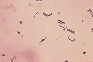
Kristal asam urat adalah   pleomorfik dibanding semua kristal urin, mereka ada dalam berbagai bentuk, seperti batang, kubus ( Gambar 6.  ), mawar enam sisi, piring, rhombi, dan seperti batu asahan. Mereka sangat birefringent dan   bervariasi dalam ukuran. Kristal asam urat   larut dalam larutan alkali dan tidak larut dalam asam. Mereka biasanya tidak berwarna sampai berwarna   kuning pucat,  pink atau coklat. Kristal asam urat sering dikaitkan dengan batu ginjal, tetapi keberadaan mereka di urin  orang normal adalah sangat umum.


GAMBAR  ,6. Kristal asam urat (panah) dan sel skuamosa.  Dalam gambar, kristal urat bentuk genjang (a) dan tampak anisotropism di bawah sinar terpolarisasi (B). (mikroskop  cahaya, × 80)  

Dalam garam asam urat  mungkin membentuk kristal lain , yaitu natrium   dan   kalium urate. Hal ini dapat dilihat sebagai tidak berwarna, berbentuk kristal jarum dan spherules kecoklatan. Penambahan setetes asam asetat glasial menunjukkan hasil spheroids  

Kalsium Oksalat

Kristal kalsium oksalat   yang paling sering diamati pada  urine asam dan   netral ( Gambar 7. ). Varian yang umum   adalah bentuk dihidrat, sebuah oktahedral, kristal berwarna mirip bentuk amplop.  Kristal jenis ini ditemukan dalam   urin   normal, terutama setelah menelan asam askorbat dalam dosis besar atau makanan yang kaya akan asam oksalat seperti tomat atau asparagus.   Bentuk lainnya adalah monohidrat, berbentuk seperti halter  atau  elips tergantung pada apakah posisi datar atau miring ( Gambar.  8 ).

GAMBAR  ,7.
Kristal kalsium oksalat  , bentuk dihidrat. berbentuk persegi seperti "bintang," atau "envelope ",  penampilan yang khas. (  mikroskop cahaya, × 160.) 


GAMBAR .8,.   Kristal kalsium oksalat, bentuk monohidrat. Catatan penampilan oval ketika berbaring datar, bentuk halter ketika miring. Dari urin pasien   penyakit kuning. (  mikroskop cahaya, × 160.)

Kristal Asam Hippuric  

Kristal asam hippuric   terkait dengan pH netral. Kristal ini biasanya   tidak berwarna, prisma memanjang dengan ujung piramida,   juga bisa tipis dan berbentuk jarum. Mereka birefringent dan terkait dengan diet tinggi buah-buahan dan sayuran yang mengandung sejumlah besar asam benzoat   

Kristal  Amorf  Fosfat

Kristal   fosfat adalah kristal yang paling sering diamati terkait dengan urin alkali. Yang paling sering dijumpai adalah kristal amorf fosfat., ini tidak dapat dibedakan dari kristal amorf urat dalam urin asam.   Kristal   menghasilkan endapan putih di dasar tabung centrifuge.  .

Kristal  Triple   Fosfat

Triple fosfat (amonium-magnesium fosfat)   adalah kristal birefringent bentuknya mirip sebuah "peti mati-tertutup"  ( Gambar  9 ),  birefringent dan sangat bervariasi dalam ukuran. Kristal juga dapat ditemukan dalam urin netral dan larut dalam asam asetat.


GAMBAR  .9. kristal  Fosfat Triple  dalam urin dengan  latar belakang Gips hialin (panah)    . (  mikroskop cahaya, × 160)  

Kadang-kadang ditemukan dalam urin basa biasanya berbentuk "bintang"  

Kristal Amonium Biurate   
Kristal Amonium   biurate memiliki bentuk "duri apel"  ( Gambar 10. ) Berwarna coklat kekuningan dan sering menunjukkan striations radial atau konsentris di   pusat seperti "senjata" atau spikula. Mereka biasanya ditemukan di dalam urin dengan pH netral dan larut dalam natrium hidroksida. Mereka jarang ditemui pada urin normal. 


GAMBAR  10. kristal Amonium biurate   dalam urin.Berbentuk  "kepiting ",  spiculated kristal merupakan ciri khas dan berkaitan dengan urin alkali. (  mikroskop cahaya, × 400.) 

Kristal  Kalsium Karbonat  
kristal karbonat kalsium berbentuk spherules-halter  kecil   ditemukan dalam urin basa ( Gambar.  11 ). Karena ukurannya yang kecil, mereka sering disangka bakteri.   Bakteri tidak birefringent. Kristal-kristal larut dalam asam asetat  . 


Gambar  11) berbentuk halter kalsium karbonat. Kristal yang ditampilkan di sini dengan kristal  triple fosfat kecil   (mikroskop, × 160. 


CAST
  Didefinisikan sebagai struktur mikroskopis silinder yang terbentuk di nefron distal dan terjadi dalam urin normal  ataupun bila ada penyakit.  Protein spesifik ini berbentuk "silinder"    yang  diproduksi hanya di tubulus distal dan duktus colleductus nefron,   protein ini larut  dan membentuk pita protein tipis yang kemudian  menyatu  atau  menjadi gips. Dalam keadaan normal, hanya ada dua varietas gips muncul dalam sedimen urin: hialin gips dan granular cast. Setiap bentuk baru harus dianggap "abnormal" dan terkait dengan penyakit ginjal metabolik umum atau intrinsik. Setiap jenis   dibahas secara terpisah.  

 TABEL  .3. KLASIFIKASI  CAST

Aselular                        Cellular  

Normal                         Normal
Hening                          Tak satupun
Granular                       Tak satupun
Abnormal                     Abnormal
Hening                          Sel darah merah
Granular                       Leukosit
Lunak                           Epitel (RTE)
Pigmen                         Lemak / lemak tubuh oval
Berlemak                      Bakteri / jamur

RBC, sel-sel darah merah, WBC, sel darah putih; RTE, epitel tubular ginjal.


Pada orang normal,   sejumlah kecil hialin atau granular  satu atau dua per 10 LP (obyektif 10 x) pada urin sering ditemukan dan tidak   selalu berarti terkena penyakit ginjal.  Kedua bentuk gips memiliki indeks bias rendah dan karena itu agak sulit untuk dilihat dengan mikroskop cahaya biasa kecuali kontras ditingkatkan. Menutup diafragma iris  sambil menurunkan kondensor dan mengatur intensitas cahaya   akan menghasilkan kontras yang optimal untuk pengamatan.   Scan slide mikroskopik secara menyeluruh untuk menemukan adanya Hialin atau Granular, dan jika ditemukan, lakukan  identifikasi   dengan menggunakan lensa   40 ×.

Cast hialin

Ini adalah yang paling sering diamati dalam urin. Bentuknya yang transparan (indeks bias yang rendah) menyebabkan agak sulit untuk dilihat. Bila diteliti  tampak perimeter luar halus dan sebuah matrik yang   halus atau bergelombang ( Gambar .12. )    Sesekali butiran inklusi  mungkin ada dalam matriks, dan kadang-kadang sel satu atau dua juga mungkin terlihat. Cor mungkin memiliki bentuk  "ekor" atau titik.  
 Di masa lalu,   gip dengan ekor disebut  cylindroid, istilah ini dianggap kuno dan tidak umum digunakan saat ini ( Gambar  13. ). 


GAMBAR  .12. Hialin cast,   struktur protein bening (panah) sering ditemukan pada sedimen urin normal


GAMBAR  13. Urine cylindroid, gip hialin dengan ekor.   Cylindroid, istilah kuno.  ( mikroskop, × 160.)

  Ketika seorang pasien mengalami stres fisik atau emosional dalam 24 jam sebelumnya, ditemukannya cylindruria tidak harus dianggap patologis., jika situasi stres atau latihan fisik telah berhenti  urin kembali ke keadaan normal dalam waktu 24 hingga 48 jam     

Granular Cast

Cast ini juga dapat diamati dalam jumlah meningkat di urin jika pasien telah terlibat dalam situasi stres emosional atau telah menjalani latihan fisik berat   Dibandingkan dengan gips hialin, granular gips ditemukan dalam rasio sekitar empat hialin per satu granular. Pada penghentian stres atau latihan, jumlah butiran gips di urin kembali normal dalam waktu 24 hingga 48 jam. Alasan peningkatan produksi terkait stres atau latihan tidak diketahui. Juga tidak diketahui alasan mengapa granular gips kadang muncul dalam urin pasien pada pola makan yang kaya karbohidrat.
Granular   memiliki indeks bias lebih tinggi daripada  hialin dan karena itu lebih mudah ditemukan. Mereka juga silindris,  walaupun beberapa mungkin memiliki "ekor," dan memiliki perimeter.  Umumnya, pada orang normal, butir menutupi permukaan cor kecil dan teratur ( Gambar.  14 ). Asal-usul butiran dalam orang normal   sebagian berasal dari partikel lisosomal  intraseluler yang dikeluarkan ke dalam urin sebagai produk metabolik dari epitel tubular ginjal  . Ketika dalam aliran urin, butiran lisosomal  masuk  ke dalam matriks cast hialin   dan dengan demikian mengubah dari yang sebelumnya mulus ( cast hialin)  menjadi kasar (cast granular).

.  

GAMBAR  ,14. Granular cast   Dalam contoh yang ditunjukkan di sini (panah), butiran-butiran tidak menutupi seluruh permukaan cor tetapi relatif merata. (  mikroskop cahaya, × 160.)